Imagine a future where medical facilities can quickly produce a 3D-printed model of a patient’s blood vessels from their CT scans, assess how their blood reacts, and utilize AI to forecast their risk of stroke years in advance. This potential is being explored by researchers at the University of Sydney, who have recently pioneered a method for 3D printing blood vessels, as detailed in their publication in Advanced Materials. This innovative technology allows the production of highly accurate vessel models that replicate the complex cartography and fluid dynamics of blood flow, which could greatly enhance the study of stroke causes and contribute to the development of tailored treatments.
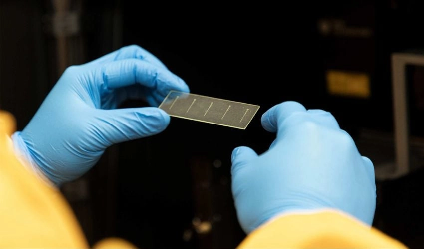
While 3D printing blood vessels is not new, this approach is distinguished by its operational efficiency. By employing CT scans from stroke patients, the team scaled down the carotid artery models to dimensions between 200 and 300 micrometers, significantly smaller than the actual 5 to 7 mm size of the artery. These miniature models were then printed on glass slides, accurately mirroring both healthy and pathological blood vessels, including common stroke-related features such as dents and irregularities. The result was a substantial reduction in printing time, from ten hours to just two.
PhD candidate Charles Zhao, from the School of Biomedical Engineering, emphasized the importance of speed and precision in stroke diagnosis, noting that clinicians often have a mere 12-hour window for decision-making post-symptom onset.
The researchers utilized digital light processing (DLP) 3D printing on glass substrates to create these so-called "patient-specific carotid artery-on-a-chip devices." This method, paired with custom clamping techniques, achieved an impressive success rate near 100%.
Once printed, the blood vessels appeared like intricate engravings on glass, but beneath this delicate surface, the researchers conducted precise blood flow simulations that mimicked the natural movement of blood. Visualizing these fluid dynamics proved to be one of the biggest challenges in the field, marking a significant milestone when accomplished.
By examining the models under a microscope, the team observed blood clot formation and the behavior of platelets—the essential elements involved in clotting that can precipitate strokes. Their findings indicated that the friction and forces exerted by blood flow against the vascular walls had a substantial effect on platelet activity. Notably, the study highlighted that areas under higher stress experienced 7 to 10 times more platelet activity.
With a "physical twin" representing the patient’s vascular system, the researchers aim to advance personalized vascular medicine. Helen Zhao, a postdoctoral digital scientist with the team, shared aspirations of integrating artificial intelligence into their biofabrication platform to create "digital twins" capable of predicting stroke incidents before they occur, thus shifting the paradigm from reactive to proactive healthcare.
This method marks a notable advancement in organ-on-a-chip technology appropriate for patient-specific applications. For further details, you can read the full study here.
What are your thoughts on utilizing 3D-printed blood vessels for stroke research? Share your thoughts in the comments or connect with us on LinkedIn and Facebook.